Cardiac Monitor
Contents
Procedure Guidelines
9.06 CARDIAC MONITOR
FAST PATCH METHOD
INDICATIONS:
Determination and monitoring of cardiac rhythms with anticipation of defibrillation.
PROCEDURE:
- Remove clothing from patient's chest.
- Apply Fast Patch pads:
- Apply PAD and STERNUM wire to upper sternum slightly toward right shoulder.
- Apply PAD and APEX wire to the anterior (mid-axillary) line below the nipple.
- Ensure the paddles are in FAST PATCH adapter with the paddles in the proper side.
- Ensure the monitor is in the PADDLES mode in the lead selection.
THREE LEAD METHOD
INDICATIONS:
Determination and monitoring of cardiac rhythms.
PROCEDURE:
- Remove clothing from area electrodes will be placed.
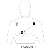
- Apply wires to electrodes.
- Place on patient as illustrated for selection lead.
INDICATIONS:
Any patient greater than 35 years of age with any of the following signs or symptoms:
- Chest pain
- Dyspnea
- Palpitations
- Weakness
- Diaphoresis
- Pre-syncope, syncope
- Nausea, vomiting
- Stroke
- Dysrythmias
- Post-resuscitation
- Pre and post-cardioversion
- During PEA to help identify the cause
- For Confirmation of Asystole
- Upper torso pain (above the umbilicus), ex. extremities
- Trauma to the Upper Torso
- Suspected Electrolyte Disturbances (e.g. DKA, dehydration, renal failure/dialysis, toxic ingestions, etc...)
PROCEDURE:
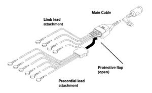
Insert the limb lead and the precordial lead attachment into the main cable as shown below:
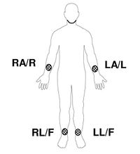
Limb Lead Electrode Sites
When acquiring a 12-Lead ECG, the limb lead electrodes are typically placed on the wrists and the ankles as illustrated below. In fact the limb lead electrodes can be placed anywhere along the limbs. However, do not place the leads on the torso when acquiring a 12-Lead ECG or you will record a non-standard report.
| AHA Labels | IEC Labels | |||
|---|---|---|---|---|
| RA | Right Arm | R | Right | |
| LA | Left Arm | L | Left | |
| RL | Right Leg | N | Negative | |
| LL | Left Leg | F | Foot | |
Precordial Lead Electrode Sites
The six precordial (chest) leads are placed on specific locations on the chest. Proper placement is important for accurate diagnosis and should be identified as shown below:
| Lead | Location |
|---|---|
| V1 | Fourth intercostal space to the right of the sternum |
| V2 | Fourth intercostal space to the left of the sternum |
| V3 | Directly between leads V2 and V4 |
| V4 | Fifth intercostal space at midclavicular line |
| V5 | Level with V4 at left anterior axillary line |
| V6 | Level with V5 at left midaxillary line. (Midpoint of armpit) |
Locating the V1 position (fourth intercostal space) is critically important because it is the reference point for locating the placement of the remaining V leads. To locate the V1 position:
- Place your finger at the notch in the top of the sternum.
- Move your finger slowly downward about 1.5 inches until you feel a slight horizontal ridge or elevation. This is the “angle of Louis” where the manubrium joins the body of the sternum.
- Locate the second intercostal space on the right side, lateral to and just below the angle of Louis.
- Move your finger down two more intercostal spaces to the fourth intercostal space, which is the V1 position.
Other important considerations:
- When placing electrodes on female patients, always place leads V3 - V6 under the breast rather than on the breast.
- Never use the nipples as reference points for locating the electrodes for men or women patients because nipple locations may vary widely.
The monitor acquires 10 seconds of ECG data for each 12-Lead ECG requested. If the monitor detects signal noise while acquiring data (such as patient movement or disconnected electrode), the monitor displays the message WAITING FOR GOOD DATA.


